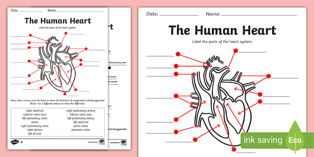
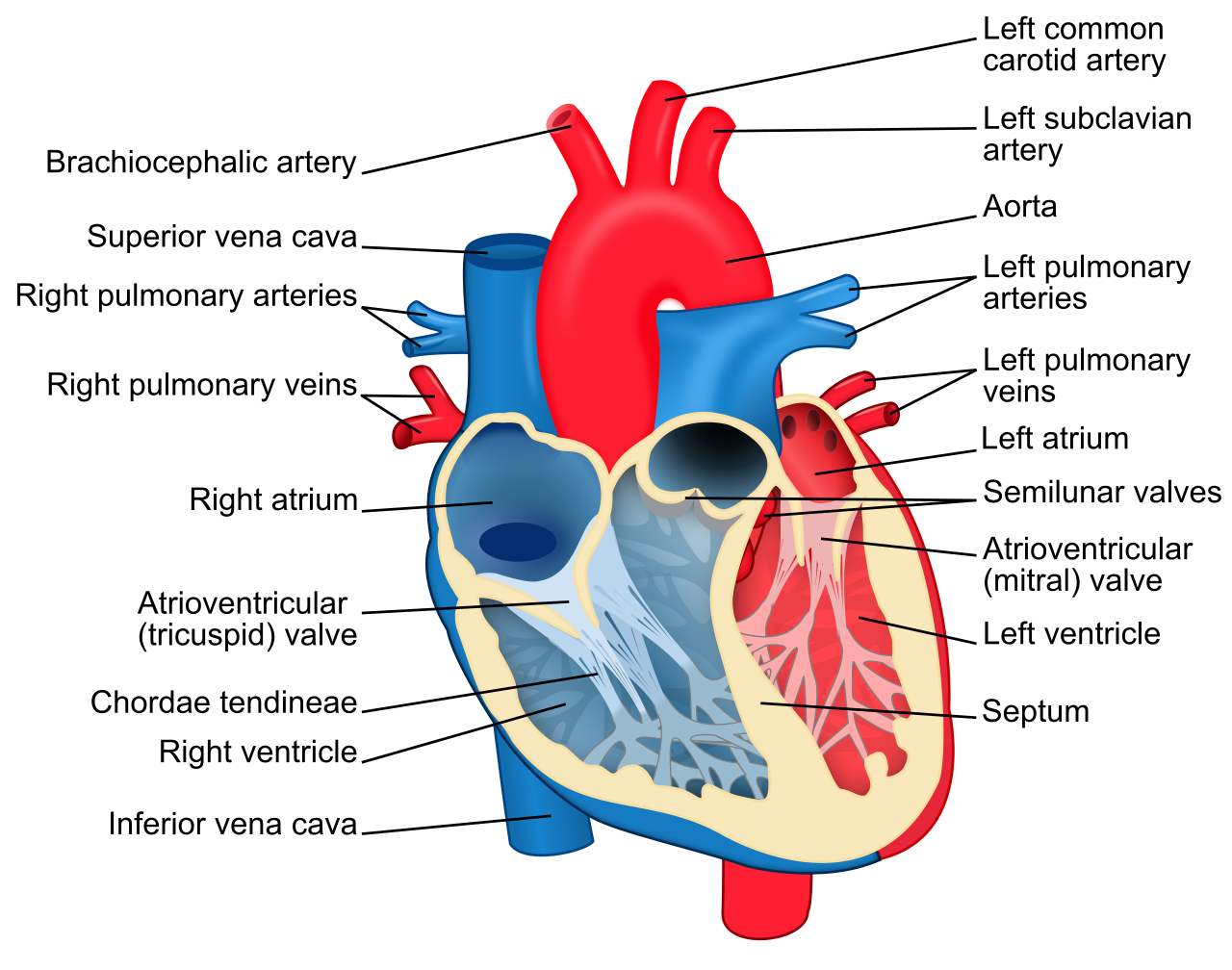
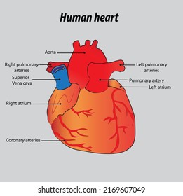
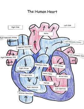
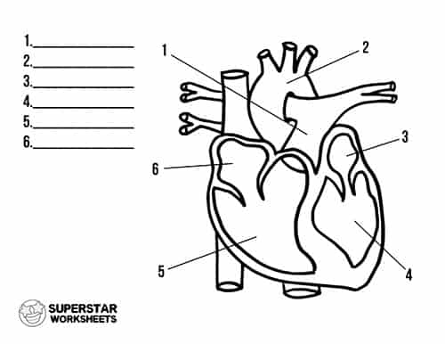
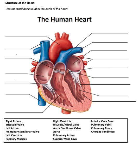
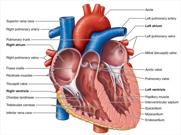
45 the human heart and its labels
File : Diagram of the human heart (cropped).svg - Wikimedia 24.10.2022 · Add Inferior vena cava and pericardium labels: 18:08, 14 August 2018: 656 × 631 (209 KB) Jmarchn (talk | contribs) Add pericardium. Add papillary muscles and chordae tendinae. Add cardiac skeleton. Inferior vena cava more wide. Add aorta in bottom. Add source veins of superior vena cava. Brachiocephalic trunk more wide and separated. Added shadows. Left … Human heart: Anatomy, function & facts | Live Science The human heart has four chambers: two upper chambers (the atria) and two lower ones (the ventricles), according to the National Institutes of Health. The right atrium and right ventricle...
› heart › picture-of-the-heartHuman Heart (Anatomy): Diagram, Function, Chambers, Location ... Cardiomyopathy: A disease of heart muscle in which the heart is abnormally enlarged, thickened, and/or stiffened. As a result, the heart's ability to pump blood is weakened. As a result, the heart ...
The human heart and its labels
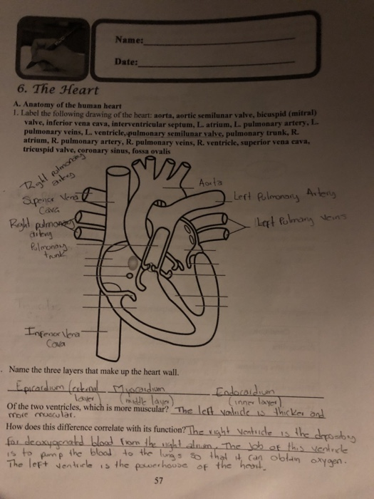
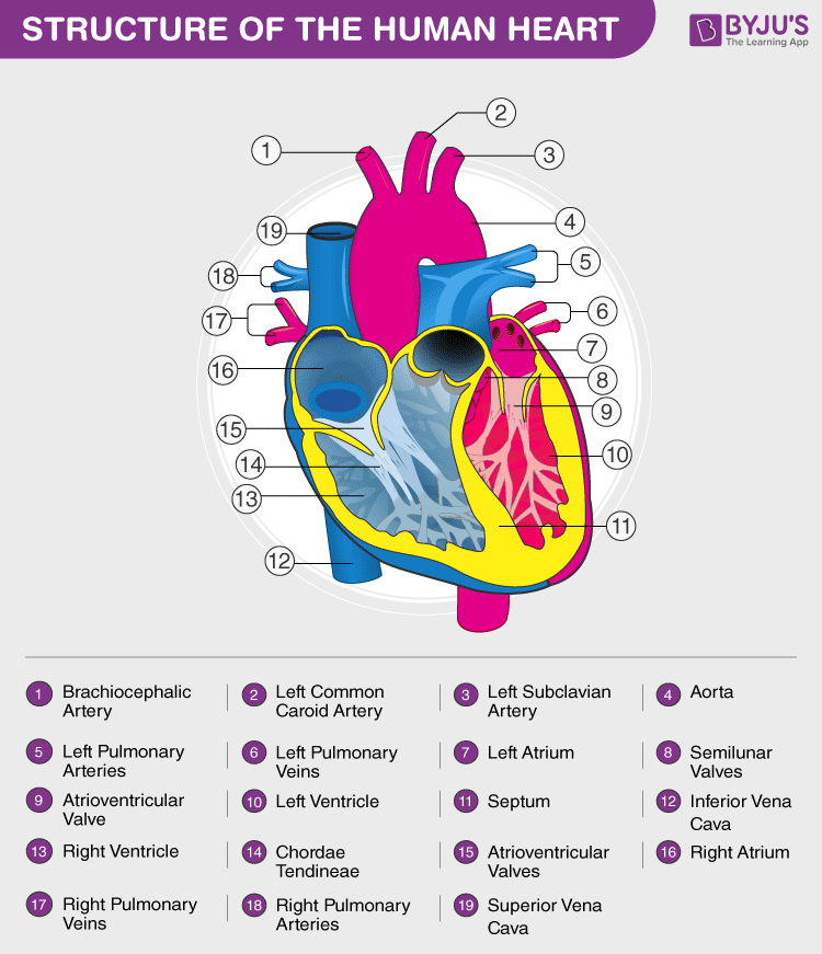
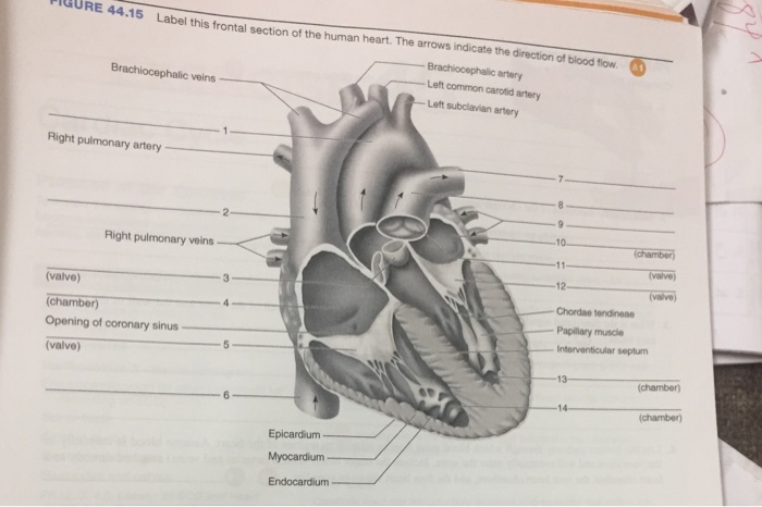
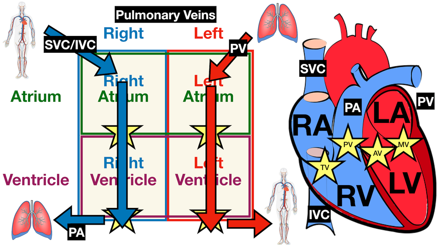
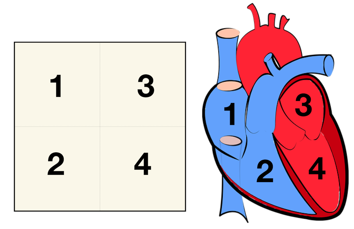
A Labeled Diagram of the Human Heart You Really Need to See The human heart, comprises four chambers: right atrium, left atrium, right ventricle and left ventricle. The two upper chambers are called the left and the right atria, and the two lower chambers are known as the left and the right ventricles. The two atria and ventricles are separated from each other by a muscle wall called 'septum'. Diagram of Human Heart and Blood Circulation in It Exterior of the Human Heart A heart diagram labeled will provide plenty of information about the structure of your heart, including the wall of your heart. The wall of the heart has three different layers, such as the Myocardium, the Epicardium, and the Endocardium. Here's more about these three layers. Epicardium Heart Diagram with Labels and Detailed Explanation - BYJUS Diagram of Heart. The human heart is the most crucial organ of the human body. It pumps blood from the heart to different parts of the body and back to the heart. The most common heart attack symptoms or warning signs are chest pain, breathlessness, nausea, sweating etc. The diagram of heart is beneficial for Class 10 and 12 and is frequently ...
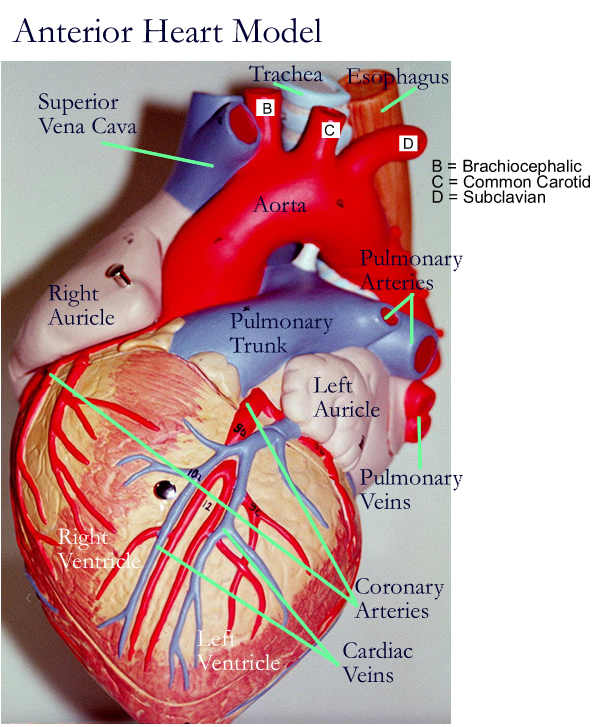
The human heart and its labels. HOW TO DRAW HUMAN HEART IN VERY EASY STEP - YouTube This is my second video on the Human heart based on general and previous knowledge which we have been reading for years. please go step by step as I am teach... How to draw & label the Human Heart || The Human Heart How to draw & label the Human Heart || The Human Heart||#educationalvideos #tashiding Human Heart Models | Heart Anatomy Models | Vitality Medical The heart model with labels is hand-painted with vivid colors to illustrate the papillary muscles, heart valves, and adjacent structures. Filter by Sort By 4 Items Magnetic Heart Model, Life Size, 5 Part G01 $394.05 View Details Classic Heart Model $81.03 View Details Human Heart Model $450.66 - $566.36 View Details heart | Structure, Function, Diagram, Anatomy, & Facts heart, organ that serves as a pump to circulate the blood. It may be a straight tube, as in spiders and annelid worms, or a somewhat more elaborate structure with one or more receiving chambers (atria) and a main pumping chamber (ventricle), as in mollusks. In fishes the heart is a folded tube, with three or four enlarged areas that correspond to the chambers in the mammalian heart. In animals ...
A Diagram of the Heart and Its Functioning Explained in Detail Human heart is covered by a double layered structure which is known as pericardium. The outer layer is associated with the major blood vessels whereas the inner layer is attached to the cardiac muscles. These layers are separated by a pericardial fluid. This covering is like a membrane which holds all the parts of the heart. Chambers Human Heart - Diagram and Anatomy of the Heart - Innerbody The heart contains 4 chambers: the right atrium, left atrium, right ventricle, and left ventricle. The atria are smaller than the ventricles and have thinner, less muscular walls than the ventricles. The atria act as receiving chambers for blood, so they are connected to the veins that carry blood to the heart. Heart: Anatomy and Function - Cleveland Clinic Heart. Your heart is the main organ of your cardiovascular system, a network of blood vessels that pumps blood throughout your body. It also works with other body systems to control your heart rate and blood pressure. Your family history, personal health history and lifestyle all affect how well your heart works. Appointments 800.659.7822. Human Heart - Anatomy, Functions and Facts about Heart - BYJUS The human heart is divided into four chambers, namely two ventricles and two atria. The ventricles are the chambers that pump blood and atrium are the chambers that receive the blood. Among which, the right atrium and ventricle make up the "right portion of the heart", and the left atrium and ventricle make up the "left portion of the heart." 5.
Human Heart Labeling Teaching Resources | Teachers Pay Teachers Human Heart Parts and Blood Flow Labeling Worksheets - Diagram/Graphic Organizer by TechCheck Lessons 4.6 (22) $2.25 Zip This resource contains 2 worksheets for students to (1) label the parts of the human heart and (2) Fill in a flowchart tracing the path of blood flowing though the circulatory system. Answer keys included. › news › indexNews Headlines | Today's UK & World News | Daily Mail Online Oct 28, 2022 · All the latest breaking UK and world news with in-depth comment and analysis, pictures and videos from MailOnline and the Daily Mail. Parts Of The Human Heart | Science Trends The parts of the human heart can be broken down into four chambers, muscular walls, vessels, and a conductive system. The two upper chambers are called the atria, with lower parts called ventricles. These all work together to make up the vital function of your heart. Everybody knows that the human heart is the essential organ in our bodies. › laws-regs › regulations1910.1030 - Bloodborne pathogens. | Occupational Safety and ... According to the concept of Universal Precautions, all human blood and certain human body fluids are treated as if known to be infectious for HIV, HBV, and other bloodborne pathogens. Work Practice Controls means controls that reduce the likelihood of exposure by altering the manner in which a task is performed (e.g., prohibiting recapping of ...
13 parts of the human heart (and its functions) - LORECENTRAL Its opening (generated by the systole of the atrium) causes blood to travel between both regions. 3. Left Ventricle. Another major part of the heart. The left ventricle receives oxygen-rich blood from the left atrium and sends it to the rest of the body through the aortic artery. 4. Aortic sigmoid valve.
Human Heart Diagram Labeled | Science Trends Human Heart Diagram Labeled Daniel Nelson 1, January 2019 | Last Updated: 3, March 2020 The human heart is an organ responsible for pumping blood through the body, moving the blood (which carries valuable oxygen) to all the tissues in the body. Without the heart, the tissues couldn't get the oxygen they need and would die.
Heart Diagram with Labels and Detailed Explanation - BYJUS Diagram of Heart. The human heart is the most crucial organ of the human body. It pumps blood from the heart to different parts of the body and back to the heart. The most common heart attack symptoms or warning signs are chest pain, breathlessness, nausea, sweating etc. The diagram of heart is beneficial for Class 10 and 12 and is frequently ...
Diagram of Human Heart and Blood Circulation in It Exterior of the Human Heart A heart diagram labeled will provide plenty of information about the structure of your heart, including the wall of your heart. The wall of the heart has three different layers, such as the Myocardium, the Epicardium, and the Endocardium. Here's more about these three layers. Epicardium
A Labeled Diagram of the Human Heart You Really Need to See The human heart, comprises four chambers: right atrium, left atrium, right ventricle and left ventricle. The two upper chambers are called the left and the right atria, and the two lower chambers are known as the left and the right ventricles. The two atria and ventricles are separated from each other by a muscle wall called 'septum'.


















Post a Comment for "45 the human heart and its labels"