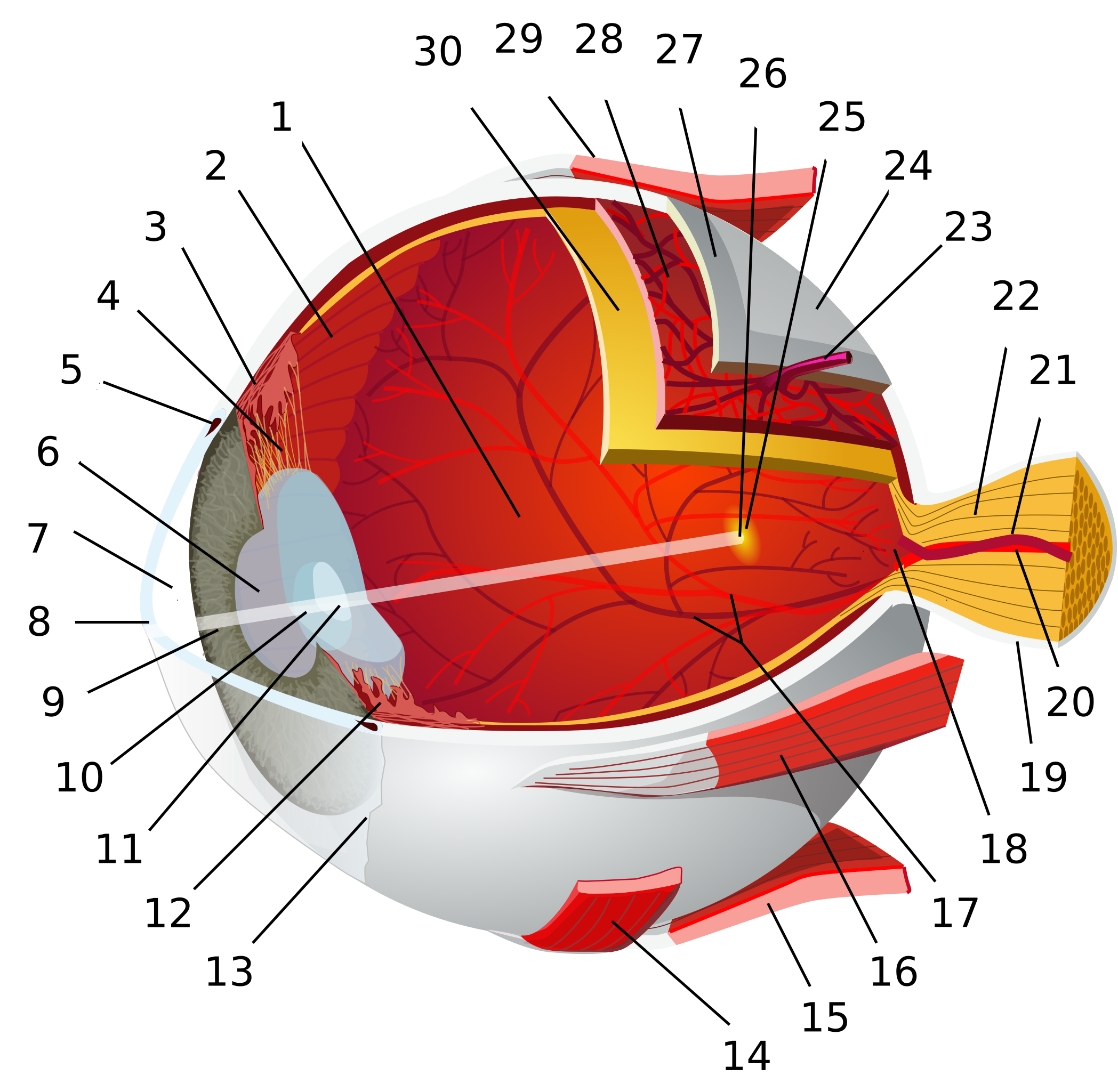
43 eye diagram and labels
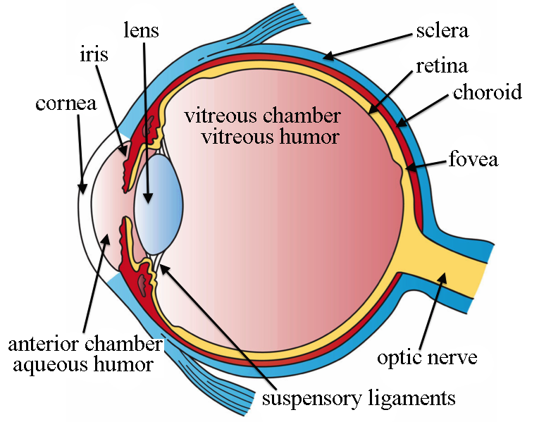
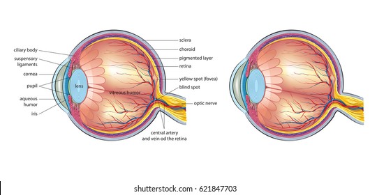
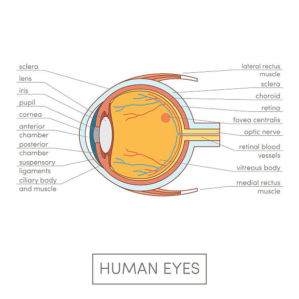
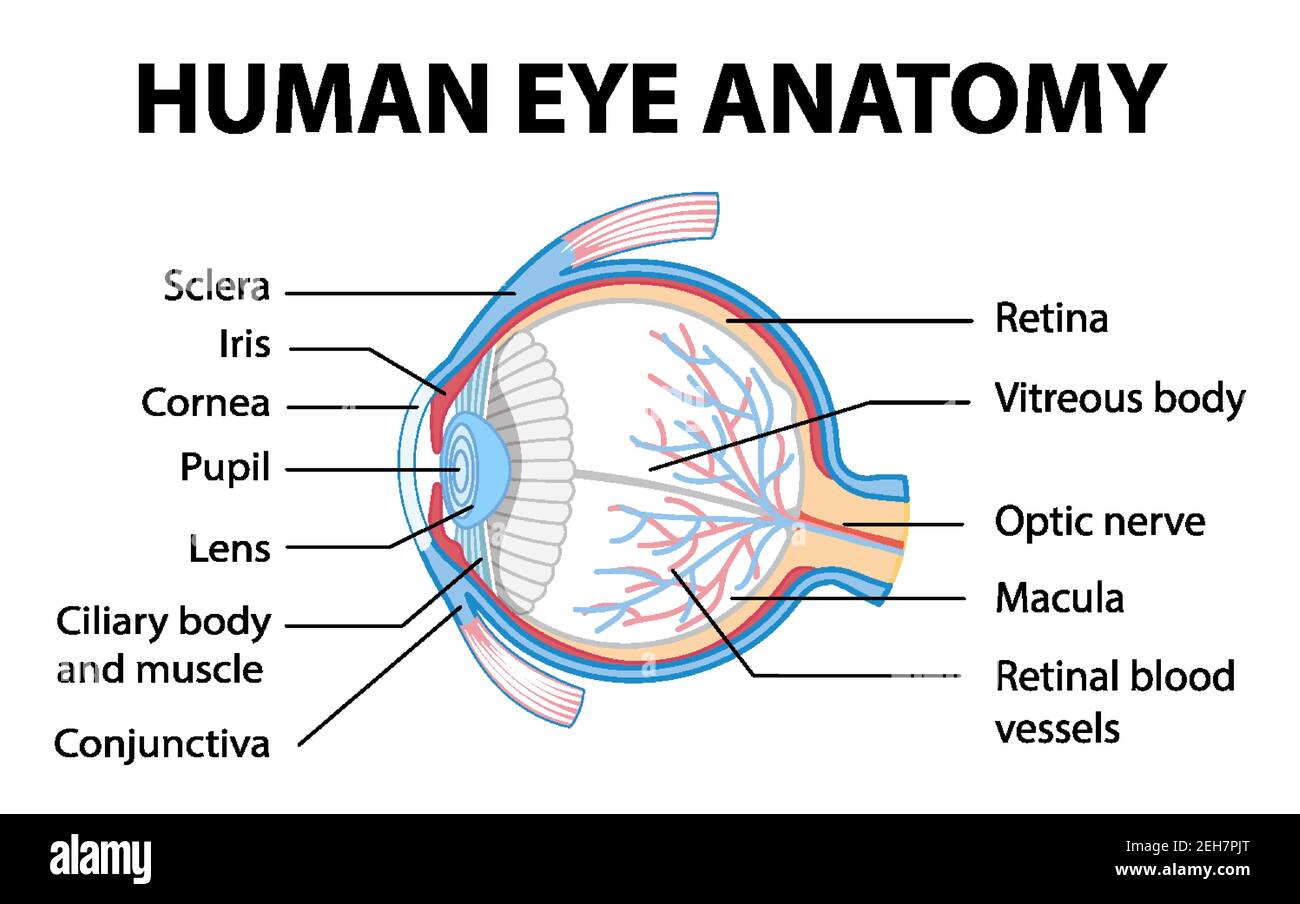
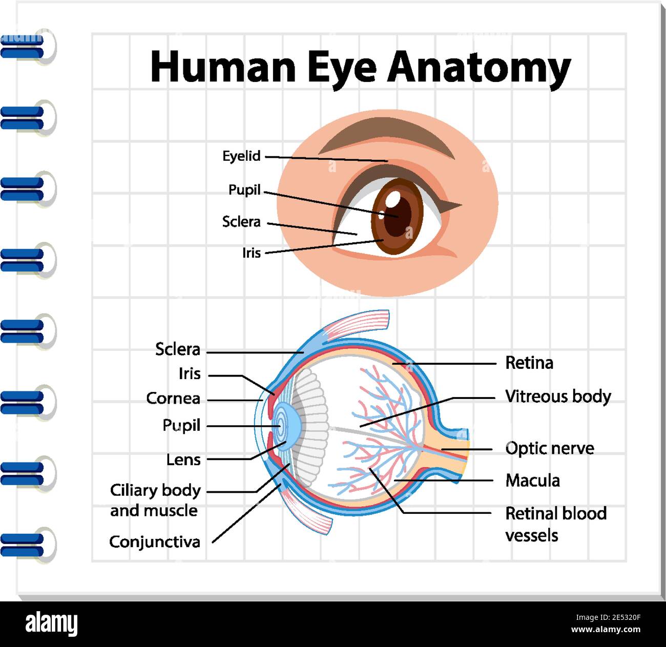
eye labeling Diagram | Quizlet It is the first structure to refract (bend) light that enters the eye. sclera Tough white out covering of the eyeball choroid Middle layer of the eye (between the retina and the sclera) that contains the blood vessels that nourish the eye and cornea iris colored layer that dilates and constricts to allow in more or less light ciliary body › conditions › pink-eye-medicinePink Eye Medicine: OTC and Prescribed - All About Vision Aug 21, 2020 · Individual drug labels can provide additional information on potential side effects or complications. When needed, steroid drops can cause the pressure inside your eye to increase. Your eye doctor may recommend monitoring your eye pressure, especially if you have or are susceptible to glaucoma.
Eye Diagram Teaching Resources | Teachers Pay Teachers The Human Eye Overview Reading Comprehension and Diagram Worksheet. by. Teaching to the Middle. 4.7. (65) $1.50. Zip. This passage briefly describes the human eye (900-1000 Lexile). 14 questions (matching and multiple choice) assess students' understanding. Students label a diagram of 6 parts of the eye.
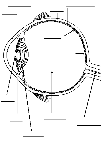
Eye diagram and labels
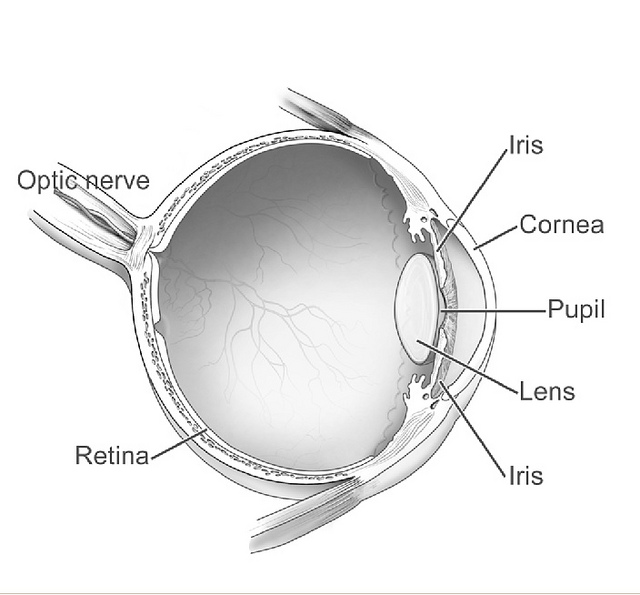
human eye diagram with labels eye diagram human labels without eyeball labeled clipart ks3 label iris physics gcse eyes body draw cyberphysics cornea lens functions. Human Skeleton Back No Text No Color Clip Art At Clker.com - Vector . skeleton human posterior clip text vector clker svg. Eye diagram labeled - Healthiack Eye diagram labeled This summary post is displaying Eye diagram labeled … The eyes are responsible for our sense of sight or vision. Any disruption of the anatomy and physiology of the eyes and their supporting structures can cause vision impairment. Some of the ocular disorders or medical conditions affecting the eyes include the following: PDF Parts of the Eye - National Institutes of Health Eye Diagram Handout Author: National Eye Health Education Program of the National Eye Institute, National Institutes of Health Subject: Handout illustrating parts of the eye Keywords: parts of the eye, eye diagram, vitreous gel, iris, cornea, pupil, lens, optic nerve, macula, retina Created Date: 12/16/2011 12:39:09 PM
Eye diagram and labels. Eye Diagram Quiz - ProProfs Quiz Try this amazing Eye Diagram Quiz quiz which has been attempted 5391 times by avid quiz takers. Also explore over 72 similar quizzes in this category. Take Quizzes. Animal; Nutrition; ... Can you label the parts of the eye in the quiz below? Give it a try and evaluate yourself. The eye has many important parts, each with different functions ... en.wikipedia.org › wiki › Graphical_user_interfaceGraphical user interface - Wikipedia The GUI (/ ˌ dʒ iː juː ˈ aɪ / JEE-yoo-EYE or / ˈ ɡ uː i / GOO-ee), graphical user interface, is a form of user interface that allows users to interact with electronic devices through graphical icons and audio indicator such as primary notation, instead of text-based UIs, typed command labels or text navigation. PDF Eye Anatomy Handout - National Institutes of Health Eye Anatomy Handout Author: National Eye Institute , National Eye Health Education Program Subject: Diabetes and Healthy Eyes Toolkit and Website Keywords: Eye anatomy, eye diagram, cornea, iris, lens, macula, optic nerve, pupil, retina, vitrous gel, diabetic eye disease. Created Date: 6/27/2012 11:57:40 AM Labelling the eye — Science Learning Hub In this interactive, you can label parts of the human eye. Use your mouse or finger to hover over a box to highlight the part to be named. Drag and drop the text labels onto the boxes next to the eye diagram If you want to redo an answer, click on the box and the answer will go back to the top so you can move it to another box.
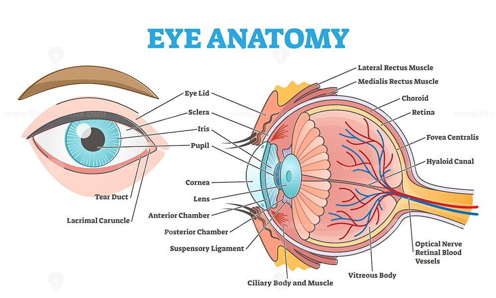
Anatomy of the eye: Quizzes and diagrams | Kenhub One of our favorite ways to get to grips with all of the parts of the eye is by utilizing labeled diagrams. On a diagram of the eye, we can see all of the relevant structures together on one image. This helps us to understand how each one is situated and related to the other. Labeled diagram of the eye Labeled Eye Diagram | Eye anatomy diagram, Eye anatomy, Diagram of the eye Eye Structure. Iridology. Simple eye diagram | Easy eye diagram | Labeled eye diagram We provide you Simple eye diagram and easy eye diagram from exam point of view. Also labeled eye diagram and anatomy of eye and human eye structure for better understanding. Human eye diagram and functions with diagram of human eye with labelling. Rudyard.org | Eye anatomy diagram, Eye anatomy, Diagram of the eye Color in the diagram as you learn what parts make up an eye. A biology resource site for teachers and students which includes lesson plans, student handouts, powerpoint presentations and laboratory investigations. Designers need to be aware that humans have common processing errors. Labeled Eye Diagram | Science Trends The human eye is composed of many different parts that work together to interpret the world around us. What you want to interpret as a major part of the human eye is somewhat up to the individual, but in general there are seven parts of the human eye: the cornea, the pupil, the iris, the lens, the vitreous humor, the retina, and the sclera. Let's take a closer look at each of these ...
Diagram of the Eye - Lions Eye Institute Instructions Click the parts of the eye to see a description for each. Hover the diagram to zoom. Need any help? If you would like to know more about us, or want to make an appointment, please don't hesitate to get in touch. (08) 9381 0777 carecentre@lei.org.au Request an appointment Customer Care Centre (08) 9381 0777 Eye labeling Diagram | Quizlet Iris a ring of muscle tissue that forms the colored portion of the eye around the pupil and controls the size of the pupil opening Cornea The clear tissue that covers the front of the eye Posterior Compartment filled with vitreous humor Pupil opening in the center of the iris Susponsory Ligament Allows the eye to move up and down vickyttoriaa Anatomy of the Eye Diagrams for Coloring/Labeling, with Reference and ... This printable contains 13 clear and simple cross sectional diagrams of the human eye. They photocopy well and are great for use as a labeling and coloring exercise for your students. The core eye anatomy diagram, designed as the labeling exercise, has a fully colored and labeled reference chart to go with it. › WAI › test-evaluateEasy Checks – A First Review of Web Accessibility Labels. Check that every form control has a label associated with it using 'label', 'for', and 'id', as shown in the labels checks below. (This is best practice in most cases, though not a requirement because a form control label can be associated in other ways.) Check that the labels are positioned correctly.
› report-covid19-resultReport a COVID-19 rapid lateral flow test result - GOV.UK Use this service to report your result to the NHS after using a rapid lateral flow test kit to check if you’re infectious with coronavirus (COVID-19). You cannot use this service to report ...
The Eye Diagram: What is it and why is it used? The eye diagram is used primarily to look at digital signals for the purpose of recognizing the effects of distortion and finding its source. To demonstrate using a Tektronix MDO3104 oscilloscope, we connect the AFG output on the back panel to an analog input channel on the front panel and press AFG so a sine wave displays. Then we press Acquire.
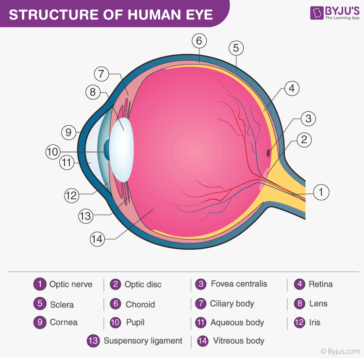
Structure and Functions of Human Eye with labelled Diagram - BYJUS Structure and Functions of Human Eye with labelled Diagram Biology Biology Article Structure Of Eye Structure of the Eye The eye is one of the sensory organs of the body. In this article, we shall explore the anatomy of the eye The structure of the eye is an important topic to understand as it one of the important sensory organs in the human body.
Eye diagram basics: Reading and applying eye diagrams - EDN Eye diagrams provide instant visual data that engineers can use to check the signal integrity of a design and uncover problems early in the design process. Used in conjunction with other measurements such as bit-error rate, an eye diagram can help a designer predict performance and identify possible sources of problems. Also see :
Labelled Diagram of Human Eye, Explanation and Function - VEDANTU Labeled Diagram of Human Eye The eyes of all mammals consist of a non-image-forming photosensitive ganglion within the retina which receives light, adjusts the dimensions of the pupil, regulates the availability of melatonin hormones, and also entertains the body clock.
6,819 Human eye diagram Images, Stock Photos & Vectors | Shutterstock Find Human eye diagram stock images in HD and millions of other royalty-free stock photos, illustrations and vectors in the Shutterstock collection. Thousands of new, high-quality pictures added every day.
Eye Diagram With Labels and detailed description - BYJUS A brief description of the eye along with a well-labelled diagram is given below for reference. Well-Labelled Diagram of Eye The anterior chamber of the eye is the space between the cornea and the iris and is filled with a lubricating fluid, aqueous humour. The vascular layer of the eye, known as the choroid contains the connective tissue.
Labelling the eye — Science Learning Hub Labelling the eye. The human eye contains structures that allow it to perceive light, movement and colour differences. In this activity, students use online or paper resources to identity and label the main parts of the human eye. Citizen science. Teacher PLD.
Eye Diagram Stock Photos, Pictures & Royalty-Free Images - iStock Browse 4,222 eye diagram stock photos and images available, or search for human eye diagram or eye diagram vector to find more great stock photos and pictures. Anatomy of human eye and descriptions. Components of human eye. Illustration about Anatomy and Physiology. Human eye anatomy.
Label Parts of the Human Eye - University of Dayton Parts of the Eye. Select the correct label for each part of the eye. The image is taken from above the left eye. Click on the Score button to see how you did. Incorrect answers will be marked in red. ...
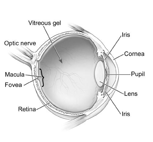
What Does the Eye Look Like? - Diagram of the Eye | Harvard Eye Associates Vitreous Gel: A thick, transparent liquid that fills the center of the eye. It is mostly water and gives the eye its form and shape. Our eyes are vital for seeing the world around us. Keep them healthy by maintaining regular vision exams. Contact Harvard Eye Associates at 949-951-2020 or harvardeye.com to schedule an appointment today.
Human eye diagram to label - simplediagram.netlify.app The figure below shows the labelled diagram of the human eye. The main parts of eye are -. Cornea Iris Pupil Ciliary muscles Eye lens retina and optical nerves which are labelled in the diagram below. The light rays coming from object enter through of eye pass through the pupil of the eye and fall on the eye.
Eye Diagram Illustrations & Vectors - Dreamstime How the eye works medical scheme poster, elegant and minimal vector illustration, eye - brain labeled structure diagram. Stylized and artistic medical design. Diagram of cataract in human eye. Illustration. Human Eye Anatomy Diagram. Eye anatomy 3d diagram infographics layout showing human eyes muscles in side view with labeling vector illustration
miro.com › templates › fishbone-diagramFishbone Diagram Template | Ishikawa Diagram Maker | Miro The Fishbone Diagram (also known as the Ishikawa diagram) is a root cause analysis tool used to identify possible causes of problems or inefficiencies in a process. The Fishbone Diagram was first created by Kaoru Ishikawa, an engineer, and professor at the University of Tokyo in 1943.
Human Eye Diagram, How The Eye Work -15 Amazing Facts of Eye FACT 1 Iris scanning is more secure than fingerprints because our iris has 256 unique characteristics and the fingerprint has just 40. FACT 2 Newborn babies don't produce tears. They only make crying sounds, but no tears come out of their crying eyes. Tears in the baby's eye start to produce when the baby is about 1-3 months old.
› en › productsElectronic Components and Parts Search | DigiKey Electronics Digi-Key is your authorized distributor with over a million in stock products from the world’s top suppliers. Rated #1 in content and design support!
en.wikipedia.org › wiki › Cerebral_cortexCerebral cortex - Wikipedia The cerebral cortex is the outer covering of the surfaces of the cerebral hemispheres and is folded into peaks called gyri, and grooves called sulci.In the human brain it is between two and three or four millimetres thick, and makes up 40 per cent of the brain's mass.
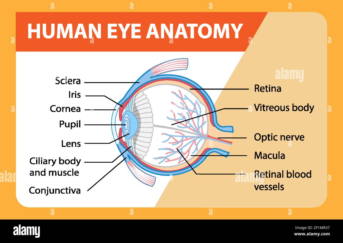
Eye Anatomy: Parts of the Eye and How We See Here is a tour of the eye starting from the outside, going in through the front and working to the back. Eye Anatomy: Parts of the Eye Outside the Eyeball. The eye sits in a protective bony socket called the orbit. Six extraocular muscles in the orbit are attached to the eye. These muscles move the eye up and down, side to side, and rotate the eye.
PDF Parts of the Eye - National Institutes of Health Eye Diagram Handout Author: National Eye Health Education Program of the National Eye Institute, National Institutes of Health Subject: Handout illustrating parts of the eye Keywords: parts of the eye, eye diagram, vitreous gel, iris, cornea, pupil, lens, optic nerve, macula, retina Created Date: 12/16/2011 12:39:09 PM
Eye diagram labeled - Healthiack Eye diagram labeled This summary post is displaying Eye diagram labeled … The eyes are responsible for our sense of sight or vision. Any disruption of the anatomy and physiology of the eyes and their supporting structures can cause vision impairment. Some of the ocular disorders or medical conditions affecting the eyes include the following:
human eye diagram with labels eye diagram human labels without eyeball labeled clipart ks3 label iris physics gcse eyes body draw cyberphysics cornea lens functions. Human Skeleton Back No Text No Color Clip Art At Clker.com - Vector . skeleton human posterior clip text vector clker svg.


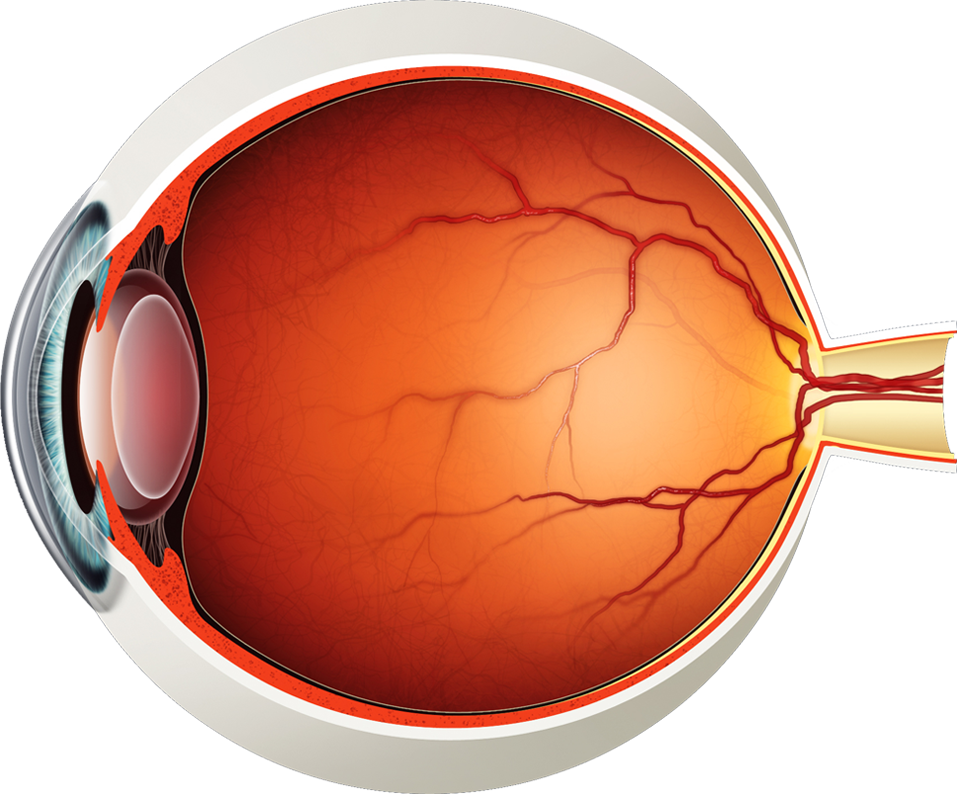
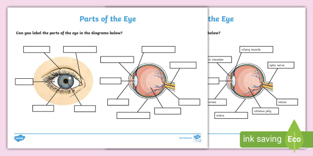


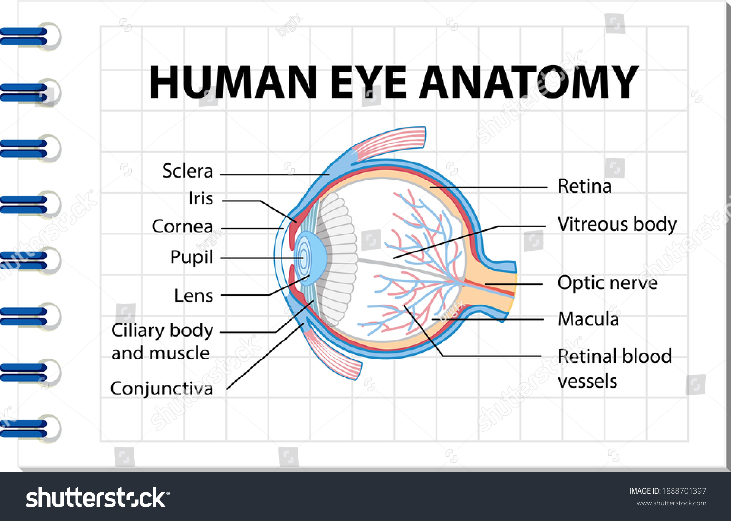

![Cross sectional diagram of human eye [1]. | Download ...](https://www.researchgate.net/publication/276541864/figure/fig1/AS:612895498964992@1523137082339/Cross-sectional-diagram-of-human-eye-1.png)


/GettyImages-695204442-b9320f82932c49bcac765167b95f4af6.jpg)







Post a Comment for "43 eye diagram and labels"